
Lameness, Severe, Cannot Support Weight on Limb Horse Side Vet Guide
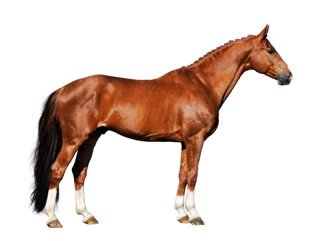
Limbs of the horse Skeletal anatomy of a horse The limbs of the horse are structures made of dozens of bones, joints, muscles, tendons, and ligaments that support the weight of the equine body.

Vitals & Anatomy Horse Side Vet Guide Horse anatomy, Anatomy, Horse care
The line leading from Eohippus to the modern horse exhibits the following evolutionary trends: increase in size, reduction in the number of hooves, loss of the footpads, lengthening of the legs, fusion of the independent bones of the lower legs, elongation of the muzzle, increase in the size and complexity of the brain, and development of crested, high-crowned teeth suited to grazing.
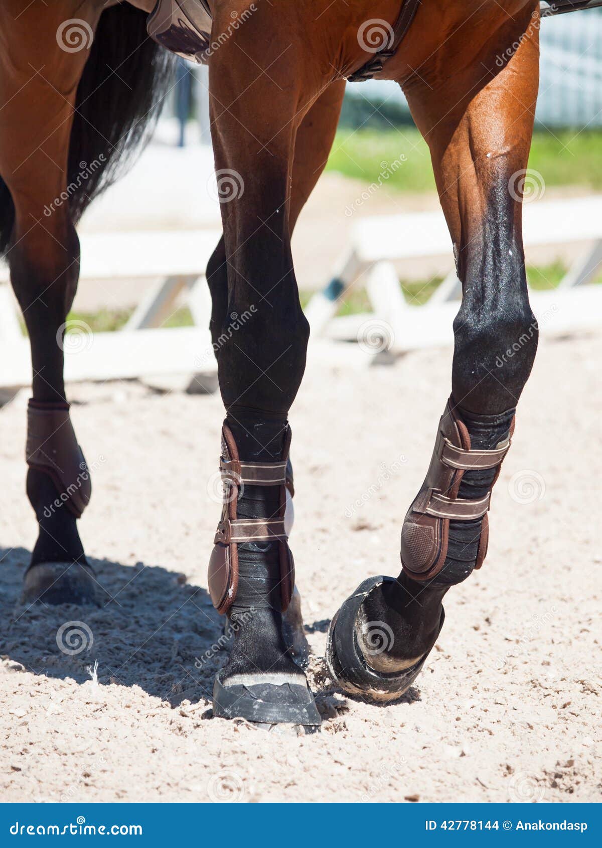
/low-section-of-horse-515042659-58d91b435f9b58468381b5ac.jpg)
White Leg Markings on Horses
Tendons attach muscle to bone. When the muscle contracts force is applied to the bone through the tendon creating force which may be either static, as in the standing horse, or result in motion. Made of carefully arranged protein fibers, primarily collagen, tendons are elastic. As long as the elastic tendon is not over stretched it recovers to.

Blood bay horse rearing on its hind legs Most Beautiful Horses, Pretty Horses, Horse Crazy
Neck: The portion of the horse's body that is between the head and shoulders. Shoulder: The upper portion of the horse's front leg. Withers: The bony ridge at the base of the neck between the shoulder blades. This ridge is created by the top portion of the thoracic vertebrae. Horses are measured at the withers.
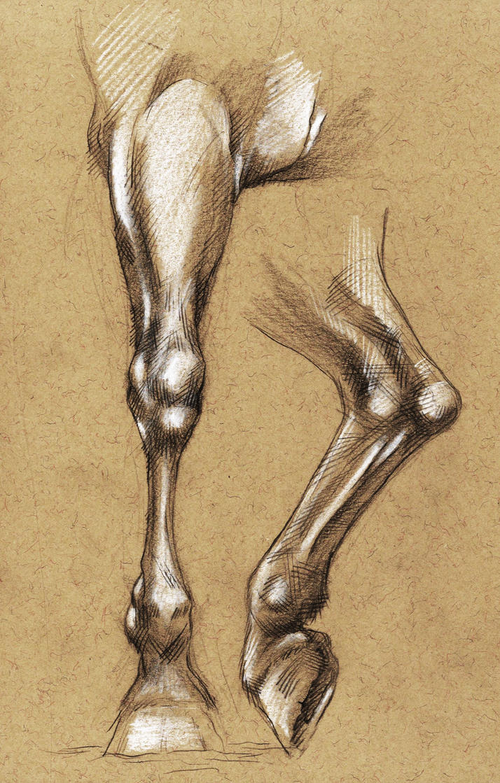
Horse leg anatomy by tirin54 on DeviantArt
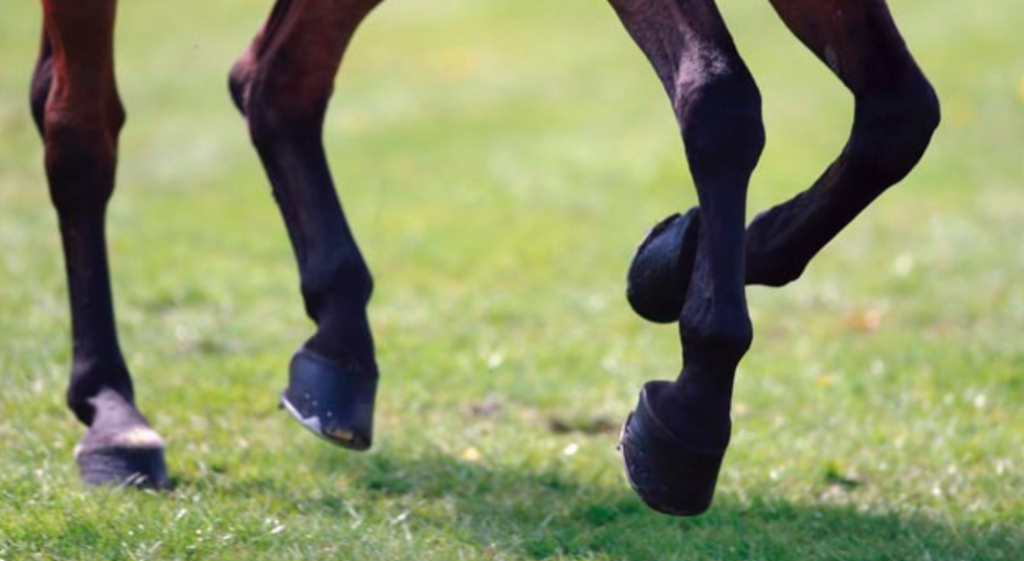
The primary function of the front legs is to support most of the horse' s weight, absorb the shock of concussion, and lift the body for the flight phase of each stride. Strongest construction consists of relatively straight legs with sturdy bone structure, big flat knees, and well-shaped fetlock joints. Straightness
:max_bytes(150000):strip_icc()/stockings-58d91d713df78c5162d10342.jpg)
White Leg Markings on Horses
Mammals Horse What's The Purpose of a Horse's Leg Chestnut? Advertisement Often called "night eyes," a horse's leg chestnut might seem unimportant now but it had a purpose years ago. Each one is unique, like a thumbprint, and grows continuously like fingernails.
horse legs Archives LubriSynHA
The horse's general form is characteristic of an animal of speed: the long leg bones pivot on pulley-like joints that restrict movement to the fore and aft, the limbs are levered to muscle masses in such a way as to provide the most efficient use of energy, and the compact body is supported permanently on the tips of the toes, allowing fuller ex.
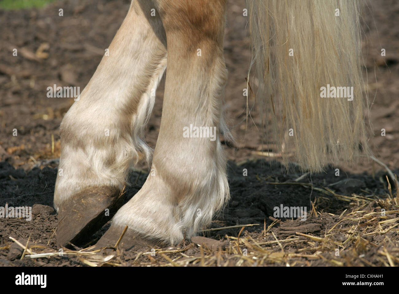
hind legs of horse Stock Photo Alamy
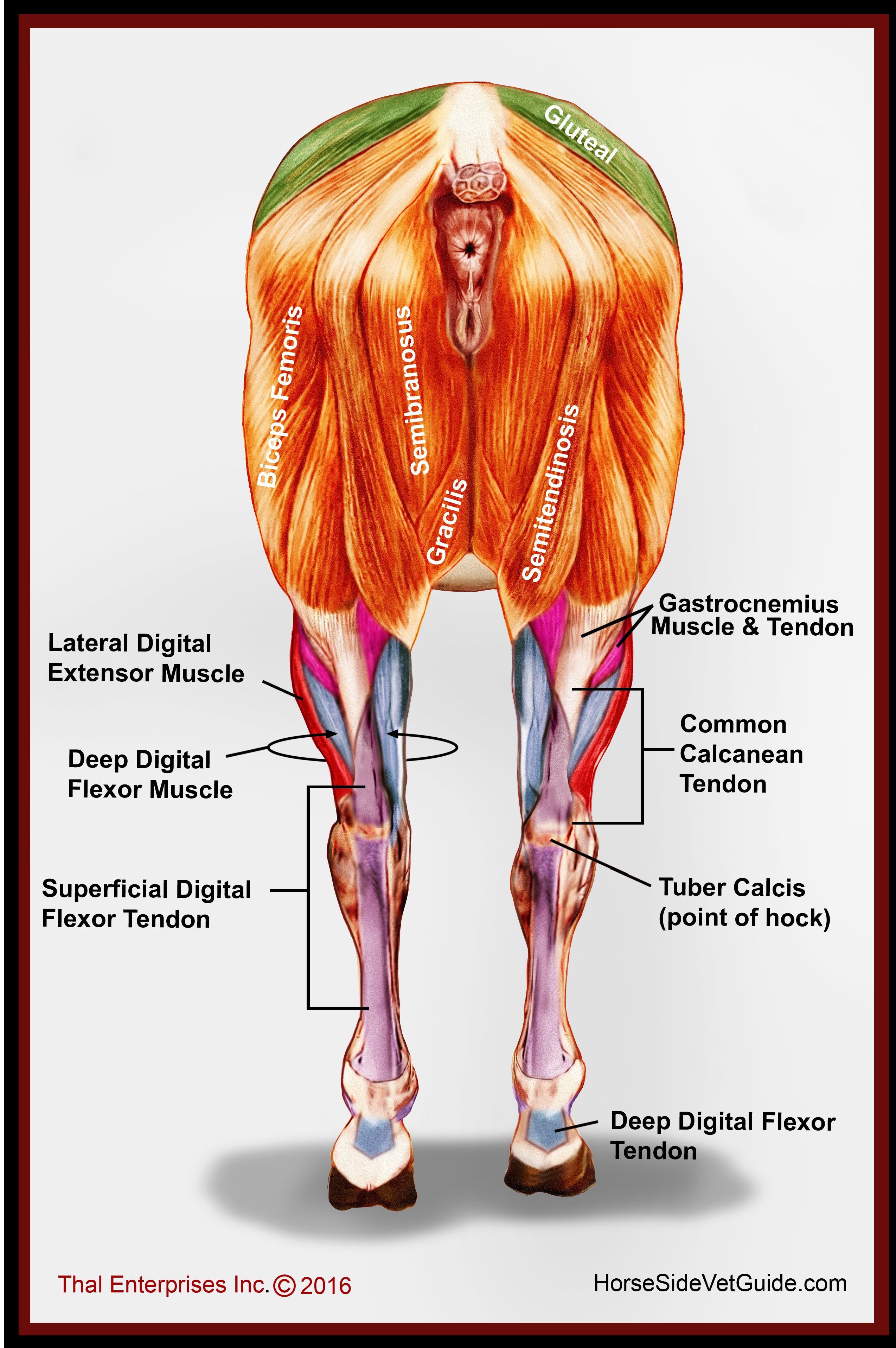
The horse leg anatomy in the rear includes the bones of the pelvis (the ilium, ischium, and pubic bones), femur, tibia, fibula, metatarsus, and phalanxes. It also includes the joints of the hip, stifles, hock, fetlock, pastern, and coffin. Hind limbs The top part of the hind limbs consists of three fused bones, called the ileum, ischium, and pubis.
Horse Legs Photograph by Carole Hinding
Chestnut (horse anatomy) The chestnut, also known as a night eye, [1] is a callosity on the body of a horse or other equine, found on the inner side of the leg above the knee on the foreleg and, if present, below the hock on the hind leg. It is believed to be a vestigial toe, and along with the ergot form the three toes of some other extinct.

Running Horse in the Desert Stock Photo Image of galloping, nature 18709594
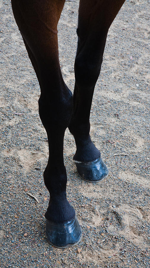
Here is a list of some of the most common conformational defects seen in the limbs of horses: Toed in: One or both legs are rotated in towards one another. Toed out: One or both legs are rotated outwards away from each other. Tied In at the Knee: The cannon bone is narrower where it ties in with the knee joint.
/GettyImages-200554179-001-5a15c93cda271500376f4638.jpg)
Hind Leg Problems in Horses Causes and Treatment
What Are The Different Parts of A Horse's Leg? Horse Leg Anatomy - Upper Hind Legs #1 - The pelvis #2 - The Femur #3 - The Stifle #4 - The Fibula and Tibia #5 - The Hock Horse Leg Anatomy - Upper Forelegs #1 - Scapular #2 - The Humerus #3 - The Elbow #4 - The Radius and Ulna #5 - The Knee Horse Leg Anatomy - Lower Legs #1 - The Cannon Bone
Database Record Viewer Horse Side Vet Guide
Since a horse's legs are made up of a finely tuned system of bones and joints, ligaments and tendons, muscles and connective tissue designed to carry a relatively heavy body, good conformation coupled with healthy limbs is extremely important for proper function. Important parts of the horse's forelimbs
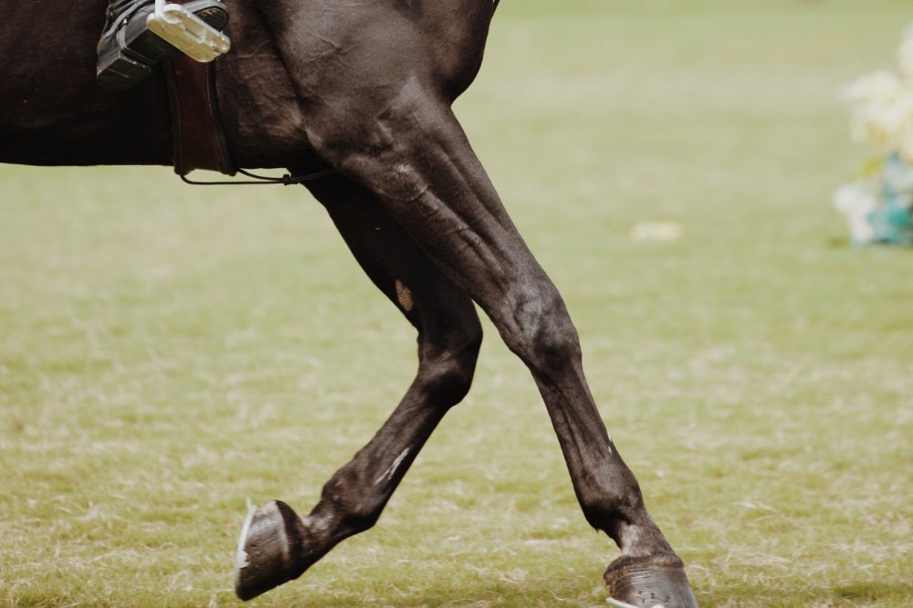
Legs of Horse in Movement. Close Up Stock Photo Image of movement, equine 42778144
Because a horse's legs are made up of a finely tuned system of bones and joints, ligaments and tendons, muscles and connective tissue designed to carry a relatively heavy body, good body conformation combined with healthy limbs is extremely important for proper function.
Horse Leg Anatomy Form and Function EquiMed Horse Health Matters
Introduction The equine hind limb is also referred to as the pelvic hind limb. When working with horses, it is important to be able to accurately assess, diagnose and manage an equine patient. To do this, a good understanding of equine anatomy is essential. Anatomy

Horse Health Helping your horse manage ringbone injury
An abnormality in a horse's movement caused by pain or reduced range of motion. It is commonly used interchangeably with the term unsoundness since a "sound" horse is one that is not lame. Lameness or Unsoundness
5 Keys to Healthy Horse Legs Draper Therapies
The hock joint is the largest joint on the horse's hind legs. The joint is made of several small bones, the most prominent being the Os Calsis which gives the hock its angular shape. The strength of the hocks is very important as this is the most active joint in the horse's hind legs. The equine hock is analogous to the human ankle.